
May 16 - 25, 2022
COURSE MATERIALS:
Below we list some relevant reviews related to microsensors and microenvironmental imaging in environmental science. PDF reprints can be downloaded as indicated
You may also want to look on the Google Scholar sites of Niels Peter Revsbech, Dirk De Beer , Ole Pedersen and Michael Kühl
A good starting point for microsensors is still the review of Revsbech&Jørgensen (1986).
Amman, R. and Kühl, M. (1998). In situ methods for assessment of microorganisms and their activities. Current Opinion in Microbiology 1: 352-358. [PDF]
Brodersen, K. E., and Kühl, M. (2022) Effects of epiphytes on the seagrass phyllosphere. Frontiers in Marine Science 9:821614. PDF
Brodersen, K. E., Kühl, M., Nielsen, D. Aa., Pedersen, O., and Larkum, A. W. D. (2018) Rhizome, Root/Sediment interactions, Aerenchyma and Internal Pressure Changes in Seagrasses. Chapter 18 in: Seagrasses of Australia. Eds. AWD Larkum, G Kendrick, PJ Ralph. Springer, pp. 393-418. PDF
De Beer, D. and Kühl, M. (2001) Interfacial processes and activities in biofilms and microbial mats. In: B. P. Boudreau and B. B. Jørgensen (eds.), The Benthic Boundary Layer. Oxford University Press, Oxford, pp.374-394. [PDF]
De Beer, D. (2000) Potentiometric microsensors for in situ measurements in aquatic environments. In: J. Buffle and G. Horvai (eds.), In situ monitoring of aquatic systems. Wiley, Chichester, pp. 161-194. [PDF]
Glud, R. N., Gundersen, J., and Ramsing, N. B. (2000) Electrochemical and optical oxygen microsensors for in situ measurements. In: J. Buffle and G. Horvai (eds.), In situ monitoring of aquatic systems. Wiley, Chichester, pp. 19-73. [PDF]
Holst, G., Klimant, I., Kohls, O., and Kühl, M. (2000) Optical microsensors and microprobes. In: M. Varney (ed.), Chemical Sensors in Oceanography. Gordon & Breach, pp. 143-188. [PDF]
Koren, K., Mosshammer, M., Scholz, V., Borisov, S. M., Holst, G., and Kühl, M. (2019) Luminescence lifetime based chemical imaging a comparison between time-domain and frequency-domain based camera systems. Analytical Chemistry 91: 32333238. PDF
Koren, K., and Kühl, M. (2018) Optical O2 sensing in aquatic systems and organisms. In: D. B. Papkovsky and R. Dmitriev (ed.), Quenched-phosphorescence detection of molecular oxygen: Applications in life sciences. Royal Society of Chemistry, Detection Science Series [PDF]
Kühl, M. and Steuckart, C. (2000) Sensors for in situ analysis of sulfide in aquatic systems In: J. Buffle and G. Horvai (eds.), In situ monitoring of aquatic systems. Wiley, Chichester, pp. 121-159. [PDF]
Kühl, M. and Revsbech. N. P. (2001) Biogeochemical microsensors for boundary layer studies. In: B. P. Boudreau and B. B. Jørgensen (eds.), The Benthic Boundary Layer. Oxford University Press, Oxford, pp. 180-210. [PDF]
Kühl, M. (2005) Optical microsensors for analysis of microbial communities. In: J. R. Leadbetter (ed.), Environmental Microbiology. Methods in Enzymology 397: 166-199. [PDF]
Kühl, M. and Polerecky, L. (2008) Functional and structural imaging of phototrophic microbial commmunities and symbioses. Aquatic Microbial Ecology 53: 99-118.[PDF]
Mosshammer, M., Brodersen, K. E., Kühl, M., and Koren K. (2019) Nanoparticle-based luminescence imaging of chemical species and temperature in aquatic systems: a review. Microchimica Acta 186: 126. PDF
Pedersen, O., Revsbech, N.P., and Shabala, S. (2020)
Microsensors in plant biology: in vivo visualization of inorganic analytes with high spatial and/or temporal resolution. Journal of Experimental Botany, 71: 39413954.
PDF
Revsbech, N. P. and Jørgensen, B. B. (1986) Microelectrodes: their use in microbial ecology. Advances in Microbial Ecology 9: 293-352. [PDF]
Revsbech, N. P., Kjær, T., Damgaard, L. R., Lorenzen, J., and Larsen, L. H. (2000) Biosensors for analysis of water, sludge and sediments with emphasis on microscale biosensors. In: J. Buffle and G. Horvai (eds.), In situ monitoring of aquatic systems. Wiley, Chichester, pp. 195-222. [PDF]
Revsbech, N. P. (2005). Analysis of microbial communities with electrochemical microsensors and microscale biosensors. In: J. R. Leadbetter (ed.), Environmental Microbiology. Methods in Enzymology 397: 147-166. [PDF]
Revsbech, N. P. (2021). Simple sensors that work in diverse natural environments: The micro-Clark sensor and biosensor family. Sensors and Actuators B: Chemical 329: 129168. PDF
Rickelt, L. F., Lichtenberg, M., Trampe, E., and Kühl, M. (2016) Fiber-optic probes for small scale measurements of scalar irradiance. Photochemistry and Photobiology 92: 331342. PDF
Santner, J., Larsen, M., Kreuzeder, A., and Glud, R. N. (2015) Two decades of chemical imaging of solutes in sediments and soils a review. Analytica Chimica Acta 878: 9-42. PDF
Scholz, V. V., Brodersen, K. E., Kühl, M., and Koren, K. (2021) Resolving chemical gradients around seagrass roots a review of available methods. Frontiers in Marine Science 8:771382. PDF
Microsensor data analysis:
Berg, P., Risgaard-Petrsen, N. and Rysgaard, S. (1998) Interpretation of measured concentration profiles in sediment porewater. Limnology and Oceanography 43: 1500-1510. [PDF] [Software] [Input file] [Manual]
[zip file with the comlete package]
PROJECTS:
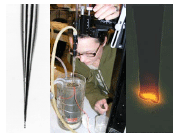
A significant part of the course involves experimental work with microsensors and related microscale techniques. The following microsensors and imaging systems will be available for experimental work:
- Electrochemical microsensors for
O2, pH, N2O, NO, H2S, Temperature
- Fiber-optic microsensors for O2 and irradiance.
- Photosynthesis quantified via variable chlorophyll fluorescence imaging
- A range of other microsensors in development/optimization
- Hyperspectral imaging system
- Optode imaging systems for O2 and pH imaging with planar and nanoparticle sensors
Given the number of participants and available supervisors we will be able to define about 4-6 project groups of 3-5 persons each. This will be done within the first 2-3 days of the course.